

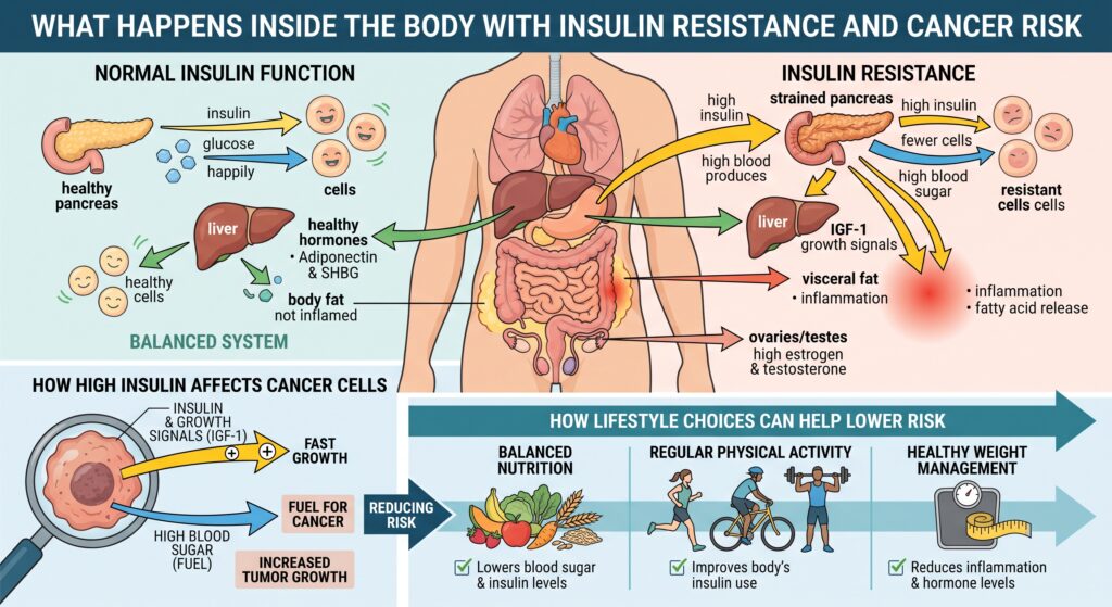
Insulin resistance and hyperinsulinemia create a systemic biochemical environment that actively promotes oncogenesis, tumor proliferation, and resistance to apoptosis. Rather than acting as a traditional mutagen, insulin resistance functions primarily as a potent metabolic driver and growth promoter across multiple cancer types, notably colorectal, breast (particularly postmenopausal estrogen-receptor-positive), endometrial, pancreatic, and liver cancers.
Key Biochemical Mechanisms
- The Insulin / IGF-1 Signaling Axis
- Receptor Cross-Activation: Chronic insulin resistance forces pancreatic beta cells to overproduce insulin to maintain glycemic control. While metabolic pathways (like glucose uptake in muscle and adipose) develop resistance, mitogenic pathways remain sensitive. High circulating insulin binds to insulin receptors IR-A isoform, often overexpressed on malignant cells) and cross-activates the Insulin-Like Growth Factor 1 (IGF-1) receptor.
- Downregulation of Binding Proteins: Hyperinsulinemia suppresses hepatic synthesis of IGFBP-1 and IGFBP-2 (IGF Binding Proteins), drastically increasing the concentration of free, bioactive IGF-1 in circulation.
- Intracellular Cascades:
- PI3K / Akt / mTOR Pathway: IGF-1 and insulin receptor activation triggers the PI3K/Akt cascade, which activates mTORC1. This stimulates ribosome biogenesis, lipid synthesis, and protein translation while shutting down autophagy and inhibiting pro-apoptotic proteins like BAD and caspase-9.
- Ras / Raf / MEK / ERK (MAPK) Pathway: Drives cell cycle progression from G to S phase by upregulating Cyclin D1.
- Substrate Availability & The Warburg Effect
- Even in the presence of oxygen, malignant cells preferentially utilize aerobic glycolysis (the Warburg effect) to convert glucose into lactate, shunting carbon intermediates into the pentose phosphate pathway for nucleotide synthesis.
- Elevated circulating glucose and free fatty acids furnish a continuous supply of substrates and ATP required to sustain rapid cell division and biomass accumulation.
- Chronic Low-Grade Inflammation & Adipokines
- Insulin resistance frequently coexists with hypertrophic, hypoxic visceral adipose tissue. Macrophages infiltrate this tissue (forming crown-like structures) and secrete pro-inflammatory cytokines: TNF-a, $IL-6, and IL-1b.
- These cytokines activate transcription factors such as NF-kappa B and STAT3, which transcriptionally upregulate cell survival factors, promote angiogenesis via $VEGF$, and facilitate immune evasion.
- Leptin vs. Adiponectin: Hyperinsulinemic states shift the adipokine balance toward elevated leptin (pro-angiogenic, anti-apoptotic) and suppressed adiponectin (an insulin-sensitizing adipokine that normally activates AMPK and inhibits tumor growth).
- Sex Hormone Dysregulation
- SHBG Suppression: High insulin directly inhibits hepatic production of Sex Hormone-Binding Globulin (SHBG).
- Elevated Free Estrogens & Androgens: Decreased SHBG elevates bioavailable, unbound estradiol and testosterone. In adipose tissue, insulin and inflammatory cytokines upregulate aromatase, accelerating the conversion of androgens to estrogens. This directly drives proliferation in hormone-sensitive tissues (endometrium and breast).
Primary Cancer Associations
| Malignancy | Primary Mechanistic Driver |
| Endometrial | Unopposed bioavailable estrogen combined with direct IRIGF-1 mitogenic stimulation. |
| Colorectal | Elevated IGF-1 suppressing epithelial apoptosis; hyperinsulinemia promoting adenoma progression. |
| Pancreatic (PDAC) | Direct exposure of local pancreatic tissue to extreme local insulin concentrations from neighboring islets, activating oncogenic KRAS pathways. |
| Hepatocellular (HCC) | Insulin resistance driving hepatic steatosis (MASLD/MASH), lipid peroxidation, oxidative stress, and cirrhosis. |
| Postmenopausal Breast | Aromatase upregulation in peripheral adipose tissue combined with low SHBG and PI3K/Akt activation. |
Therapeutic Targets & Metabolic Interventions
- AMPK Activation: Pharmacological agents like metformin and physical exercise activate AMPK, which directly inhibits mTORC1, downregulates hepatic gluconeogenesis, and reduces fasting insulin.
- SGLT2 Inhibitors & GLP-1/GIP Receptor Agonists: Improve glycemic control, reduce circulating insulin demand, and alleviate systemic visceral inflammation.
- Dietary & Lifestyle Interventions: Resistance training and aerobic exercise enhance non-insulin-dependent glucose uptake (via GLUT4 translocation), clearing glucose and lowering basal insulin secretion. Minimizing glycemic load and reducing visceral adiposity remove the upstream stimulus driving the entire signaling cascade.
When the body becomes resistant to insulin, the pancreas responds by pumping out extra insulin to help keep blood sugar under control. While insulin normally helps turn food into energy, having constantly high levels of it acts like a growth switch for cells. High insulin levels also boost other growth hormones (like IGF-1), which signal cells to divide faster and ignore the body’s natural “self-destruct” signals that normally eliminate damaged cells. At the same time, high blood sugar and excess fats provide an abundant, nonstop fuel supply that cancer cells readily consume to grow and multiply.
Beyond triggering cell growth, insulin resistance creates a hostile internal environment marked by chronic inflammation and hormonal imbalances. Carrying excess visceral fat releases inflammatory chemicals into the bloodstream, which damages tissues over time and makes it easier for tumors to form blood vessels and thrive. High insulin also disrupts sex hormones by increasing active estrogen and testosterone in the blood, significantly raising the risk for hormone – driven cancers like breast and uterine cancer, as well as colorectal and liver cancers. The encouraging takeaway is that restoring insulin sensitivity-through regular exercise, balanced nutrition, and healthy weight management – helps turn off these growth signals and lowers cancer risk.
Leave a Reply