

Can Insulin Resistance Cause the Progression of Neuropathy?
Insulin resistance significantly impairs peripheral nerve repair, and the combination of myo-inositol (MI) and D-chiro-inositol (DCI) addresses the metabolic and structural bottlenecks involved in diabetic and metabolic neuropathy.
How Insulin Resistance Impairs Nerve Repair
Insulin acts as a potent neurotrophic factor in the peripheral nervous system, directly regulating sensory neurons, axonal outgrowth, and Schwann cell survival. When insulin resistance develops, several distinct mechanisms disrupt nerve repair:
- Blunted Neurotrophic Signaling: Sensory neurons and dorsal root ganglia express insulin receptors. Under insulin-resistant conditions, downstream PI3K/Akt/mTOR signaling is blunted, suppressing protein synthesis necessary for cytoskeletal rebuilding (such as tubulin and neurofilaments) during axonal regeneration.
- Endoneurial Microvascular Hypoxia: Insulin resistance impairs endothelial nitric oxide synthase (eNOS) activity, decreasing capillary blood flow through the vasa nervorum (the tiny vessels supplying nerves). This causes local ischemia, depriving repairing axons of oxygen and essential substrates.
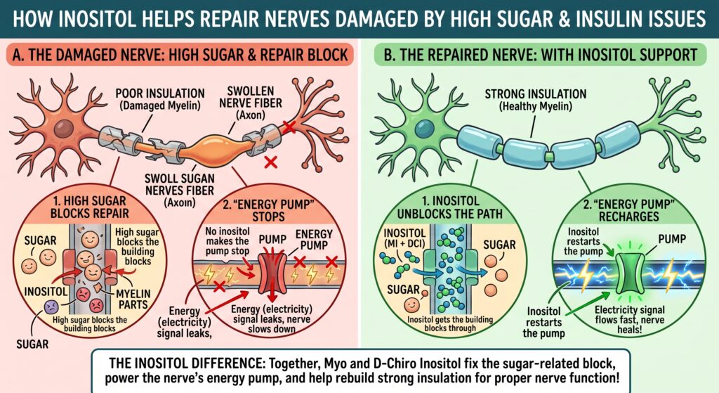
- Hyperglycemic Polyol Overload: Excess glucose shunted through the aldose reductase pathway produces sorbitol and fructose, which depletes cellular NADPH, induces oxidative stress, and competitively inhibits the sodium-myo-inositol transporter (SMIT1/2).
- Na+/K+-ATPase Dysfunction: Intracellular depletion of myo-inositol suppresses phosphoinositide turnover and Protein Kinase C (PKC) activity. This downregulates the membrane Na+/K+-ATPase pump, causing intra-axonal sodium accumulation, nodal swelling, and slowed nerve conduction velocity.
How Myo-Inositol & D-Chiro-Inositol Support Sensitivity and Nerve Formation
Myo-inositol and D-chiro-inositol work synergistically to restore both systemic insulin action and localized nerve architecture:
| Mechanism | Target Action | Impact on Nerve Repair |
| Second Messenger Function | Precursor to Inositol Phosphoglycans (IPGs) | MI-IPG enhances cellular glucose uptake (via GLUT4), while DCI-IPG stimulates glycogen synthase, lowering ambient insulin resistance. |
| Intra-Neuronal Replenishment | Bypasses competitive glucose uptake block | Floods extracellular space to restore normal intracellular inositol concentrations in peripheral axons and Schwann cells. |
| Na+/K+-ATPase Restoration | Re-establishes phosphatidylinositol pools | Reactivates the sodium-potassium pump, correcting axonal membrane potential and improving nerve conduction velocity. |
| Structural Remyelination | Provides membrane phosphoinositide backbone | Supports lipid membrane synthesis required by Schwann cells for axonal sheath formation and paranodal stabilization. |
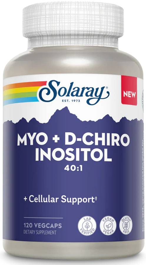
Supplementation protocols commonly leverage a 40:1 ratio of Myo-Inositol to D-Chiro-Inositol to mimic physiological plasma proportions, preventing the blunting of cellular uptake that can occur if DCI is dosed in excess.
Further on Myo D-Chiro Inositol and Nerve Health
Inositol isomers serve both as metabolic second messengers and essential structural building blocks in the peripheral nervous system. Examining their mechanisms highlights how they support axonal regeneration, maintain myelin architecture, and interface with cellular transport.
- Dual-Action Second Messengers: MI vs. DCI
Both stereoisomers act as precursors to inositol phosphoglycans (IPGs), but they control distinct arms of downstream insulin signaling:
- Myo-Inositol (MI-IPG): Stimulates glucose transporter type 4 (GLUT4) translocation to the plasma membrane and drives glucose oxidation. In neural tissue, MI is the predominant isomer (~99% of total tissue inositol). It incorporates into phosphatidylinositol 4,5-bisphosphate (PIP2), providing the precursor pool for IP3 and DAG to maintain normal axonal depolarization cascades.
- D-Chiro-Inositol (DCI-IPG): Directly activates glycogen synthase and pyruvate dehydrogenase, promoting intracellular glucose storage and disposal in peripheral tissue.
- The Epimerase Imbalance: Under conditions of chronic insulin resistance or hyperglycemia, the tissue enzyme epimerase (which converts MI to DCI) becomes dysregulated. Supplementing with both isomers bypasses this enzymatic bottleneck, improving systemic insulin sensitivity while preventing intracellular depletion in peripheral nerves.
- Transporter Dynamics & The Na+/K+-ATPase Cascade
Peripheral nerves rely heavily on active transport to maintain intracellular inositol pools:
- Competitive Uptake via SMIT: Extracellular myo-inositol enters dorsal root ganglia and Schwann cells via sodium-dependent myo-inositol transporters (SMIT1 and SMIT2). Because D-glucose and sorbitol share structural homology with inositol, elevated interstitial glucose competitively inhibits SMIT-mediated influx.
- Re-establishing the Pump: Once intracellular inositol drops, Protein Kinase C (PKC) activity collapses, resulting in deactivation of membrane Na+/K+-ATPase. Restoring saturating levels of inositol recharges the phosphoinositide cycle, reactivating the pump and restoring the electrochemical gradient necessary for proper action potential propagation.
- Schwann Cell Function and Remyelination
Schwann cells produce the lipid-rich myelin sheath that insulates large-diameter axons. Inositol plays two critical roles in Schwann cell survival and remyelination:
- Phospholipid Matrix Synthesis: Myelin is roughly 70-85% lipid by dry weight. Phosphatidylinositol and its phosphorylated derivatives are integral components of the Schwann cell membrane bilayer, stabilizing the paranodal junctions that anchor myelin loops to the axon.
- Axon-Glial Trophic Crosstalk: Dedifferentiated “repair” Schwann cells require intact PI3K/Akt signaling to transition back into myelinating phenotypes following injury. Adequate inositol availability supports the lipid kinase cascades necessary for upregulating myelin-specific transcription factors like Krox-20 (Egr2).
- Clinical Dosing Ratios & Formulations
- 40:1 Physiological Ratio: Clinical protocols typically use a 40:1 MI to DCI ratio (e.g., 2,000 mg MI to 50 mg DCI, taken 1-2 times daily). Excessively high doses of DCI relative to MI can oversaturate intestinal and cellular uptake channels, reducing the bioavailability of myo-inositol.
- Synergistic Co-Factors: Inositol is often paired with lipid-soluble metabolic agents such as Alpha-Lipoic Acid (ALA) (to suppress polyol pathway oxidative stress) or Benfotiamine (to block toxic advanced glycation end-product formation), addressing multiple neuropathic pathways simultaneously.
Summary:
Insulin resistance and chronic hyperglycemia impair peripheral nerve repair by blunting essential neurotrophic signaling, starving microvessels of oxygen, and depleting intracellular inositol pools. When excess glucose overloads the polyol pathway, it competitively blocks sodium-dependent myo-inositol transporters (SMIT1/2), precipitating a loss of membrane Na+/K+-ATPase pump activity. This transport failure leads to intra-axonal sodium retention, nodal swelling, and severely reduced nerve conduction velocities, while also depriving Schwann cells of the phosphoinositides needed to maintain and rebuild the myelin sheath.
Supplementation with a physiological 40:1 ratio of myo-inositol to D-chiro-inositol addresses both the systemic metabolic defect and local structural damage in peripheral nerves. Myo-inositol restores cellular PIP2 pools, reactivates the Na+/K+-ATPase pump, and provides the phospholipid backbone required for Schwann cell remyelination and axonal repair. Concurrently, D-chiro-inositol promotes glycogen synthesis and downstream glucose disposal, bypassing damaged epimerase pathways to improve overall insulin sensitivity and create a favorable metabolic environment for sustained neural regeneration.
Leave a Reply