

Silent Signals: How Microvascular Health Shapes Your Hearing, Balance, and Vision
When we think about circulation, we usually picture the heart and the major arteries – the multi-lane highways of the body. But branching off those major highways is a vast network of microscopic roads called capillaries. These microvessels are so narrow that red blood cells often have to travel through them in single file.
Nowhere in the human body is this micro-plumbing more vital than in your sensory organs: your inner ears and your eyes.
Your eyes and ears are among the most metabolically active tissues you possess. They never truly shut off; they constantly process photons of light and sound vibrations, transforming physical energy into electrical signals for the brain. Because they work around the clock, they depend on an uninterrupted flow of oxygen and nutrients through these tiny capillaries.
When blood vessels undergo stress – whether from fluctuating blood sugar, chronic inflammation, or oxidative damage – the smallest vessels fail first. Clinicians call this microangiopathy (literally “small blood vessel disease”). For many people, sensory changes like ringing in the ears, mild dizziness, or hazy vision are the earliest warning signs that microvascular health needs attention.
Part 1: The Inner Ear – A High-Voltage Battery Running on Tiny Capillaries
Most people assume hearing loss is simply mechanical wear-and-tear from loud concerts or aging. However, the inner ear is actually an intricate bio-electrical transducer, and its power source is purely vascular.
The inner ear consists of two main sensory chambers:
- The cochlea (the snail-shaped structure responsible for hearing).
- The semicircular canals (the loop-shaped fluid tubes responsible for balance and spatial orientation).
The Inner Ear’s Power Plant: The Stria Vascularis
Tucked inside the cochlea is a specialized band of tissue called the stria vascularis. Think of this tissue as the cochlea’s rechargeable battery pack. It maintains a very specific, electrically charged fluid called endolymph that surrounds the microscopic hair cells.
For you to hear a whisper, this fluid must maintain a precise balance of ions (mostly potassium and sodium). Keeping this electrical charge high requires massive amounts of cellular energy, which is delivered entirely by a delicate capillary bed.
What Happens When Micro-Vessels Falter?
When microvascular damage occurs:
- The “Battery” Loses Voltage: The capillaries supplying the stria vascularis narrow or become sluggish. Deprived of oxygen and fuel, the tissue cannot properly pump ions, and the electrical charge of the fluid drops.
- Hair Cells Starve: Without the proper ion balance, the fragile sensory hair cells cannot convert sound vibrations into nerve impulses. Over time, this leads to bilateral sensorineural hearing loss – gradual, permanent hearing loss affecting both ears, especially noticeable when trying to decipher speech in noisy rooms.
- The “Gain” Gets Turned Up (Tinnitus): When the brain stops receiving clean sound signals from the cochlea, it attempts to compensate by turning up its internal auditory “volume.” This phantom signal is perceived as tinnitus – constant ringing, hissing, buzzing, or clicking in the ears.
- Balance System Disruptions (Vertigo): Because the balance canals share the same fluid network, fluid and vascular imbalances can disrupt your equilibrium sensors. This results in vertigo, an unsettling sensation that the room is spinning, or a persistent feeling of being lightheaded and unsteady.
Part 2: The Eyes – When the Body’s Camera Loses Its Supply Line
Just like the inner ear, the eye requires immense vascular precision. It functions like a sophisticated camera, where clear optical elements focus light onto an ultra-sensitive digital sensor.
When microvascular stress sets in, three distinct areas of the eye – the retina, the lens, and the internal drainage channels – can become compromised.
1. Retinal Capillaries: Damage That Starts Before Diagnosis
The retina is the light-sensitive lining at the back of your eye. It has one of the highest metabolic rates of any tissue in the entire body.
Under conditions of metabolic strain (such as insulin resistance or elevated blood sugar), the walls of the retina’s micro-capillaries weaken:
- They develop tiny pouch-like bulges (microaneurysms) and may begin to leak small amounts of fluid or blood into the retinal tissue.
- Crucially, clinical research reveals that this microvascular damage often begins in prediabetes, years before an individual crosses the threshold into diagnosable Type 2 diabetes.
- Because early retinal damage rarely hurts or causes dramatic blind spots right away, it frequently goes unnoticed until significant capillary networks are blocked.
2. Early-Onset Nuclear Cataracts: A Fogged Lens
Directly behind the pupil sits the crystalline lens, which fine-tunes your focus. Unlike the retina, the lens has no direct blood supply of its own; it absorbs nutrients from the clear fluid circulating around it.
When systemic vascular and metabolic health is disrupted, the lens faces two major stresses:
- Osmotic Stress: Excess sugar molecules in the surrounding fluid pull water into the lens through osmosis. The lens fibers swell, disrupting the crystalline order that makes the lens transparent.
- Oxidative Stress: Free radicals overwhelm the eye’s natural antioxidant defenses, chemically degrading the lens proteins.
Instead of remaining crystal clear, the core of the lens (the nucleus) turns cloudy and yellowish. This accelerates early-onset nuclear cataracts, causing colors to look washed out, worsening night glare around headlights, and creating a sensation of looking through a dirty or frosted window years ahead of normal aging.
3. Glaucoma: Clogged Drains and Optic Nerve Pressure
The front part of your eye continually produces a clear fluid called aqueous humor, which must drain out at an equal rate through a microscopic mesh network (the trabecular meshwork).
When microvascular inflammation and poor blood flow impair the cells that regulate this drainage system:
- Fluid cannot escape fast enough, causing intraocular pressure (IOP) to rise.
- This elevated pressure pushes backward into the eyeball, compressing the tiny vessels that feed the optic nerve.
- Deprived of oxygenated blood, optic nerve fibers gradually die off, resulting in glaucoma – a condition that slowly eats away at peripheral vision from the outside in.
Summary: Comparing Ear and Eye Microvascular Complications
The following breakdown highlights how microvascular changes directly manifest in sensory tissues:
| Sensory Organ | Microscopic Target | What Happens Pathologically | Common Everyday Symptoms |
| Inner Ear (Cochlea) | Stria vascularis (vascular wall) | Ion transport fails; cochlear fluid loses its electrical charge | Muffled hearing in both ears, difficulty understanding conversations in crowds |
| Inner Ear (Canals) | Semicircular canal fluid system | Fluid pressure/chemistry imbalances in motion sensors | Vertigo (spinning sensations), balance instability, head tilt dizziness |
| Auditory Nerve Pathway | Sensory hair cell synapses | Brain compensates for missing input by amplifying internal static | Persistent ringing, whistling, or buzzing (tinnitus) |
| Eye (Retina) | Retinal capillaries | Micro-vessels weaken, leak fluid, or close off entirely | Blurred central vision, dark floaters, reduced low-light vision |
| Eye (Lens) | Lens structural proteins | Osmotic swelling and oxidative clumping of proteins | Glare around night lights, dull color contrast, “foggy” vision |
| Eye (Drainage System) | Trabecular meshwork & optic disc | Poor drainage elevates intraocular pressure, pinching nerve fibers | Loss of peripheral (“tunnel”) vision, subtle eye pressure |
Protecting Your Microvascular Health
Because the capillaries of the eye and ear are among the smallest in the body, they serve as an “early warning system.” Safeguarding them involves supporting overall metabolic and vascular health:
- Schedule Annual Dilated Eye Exams: An optometrist or ophthalmologist is the only specialist who can directly look at live microvessels without making an incision. Retinal imaging can spot microvascular changes years before they trigger noticeable vision loss.
- Get a Baseline Hearing Test: If you notice sudden ringing, muffling, or dizziness, do not brush it off as simple earwax or fatigue. Audiograms can detect early high-frequency loss caused by stria vascularis fatigue.
- Monitor Metabolic Health Early: Since microvascular damage begins at prediabetic levels, checking fasting glucose, HbA1c, fasting insulin, and blood pressure provides actionable insight long before irreversible sensory damage occurs.
- Target Inflammation and Oxidative Stress: Consistent cardiovascular activity, blood pressure management, and a diet rich in polyphenols and antioxidants help maintain healthy capillary lining (the endothelium) throughout the body.
The Missing Link: Insulin Resistance!
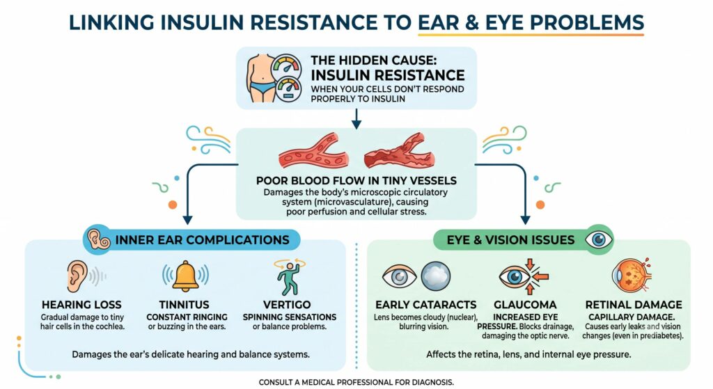
Insulin resistance is the hidden engine driving every single one of these microvascular breakdowns.
When people hear “insulin resistance,” they usually think of weight gain or full-blown Type 2 diabetes. In reality, insulin resistance damages your smallest blood vessels years – often a decade or more – before fasting blood sugar ever flags a red warning on a standard lab test.
To understand why your ears and eyes suffer first, it helps to see how insulin resistance quietly sabotages micro-circulation from the inside out.
The Domino Effect: How Insulin Resistance Damages Microvessels
Insulin’s primary job is acting like a key that unlocks your cells so they can absorb glucose for energy. When your cells become resistant to that key, two destructive things happen simultaneously:
- Insulin Levels Spike (Hyperinsulinemia): The pancreas pumps out larger and larger surges of insulin to force glucose into stubborn cells. High circulating insulin acts like an irritant inside delicate blood vessels, triggering chronic low-grade inflammation.
- Nitric Oxide Drops (Vessels Clamp Shut): Healthy capillary linings produce nitric oxide, a natural gas that tells blood vessels to relax, open wide, and let blood flow smoothly. Insulin resistance blunts nitric oxide production. As a result, microvessels chronically constrict and stiffen, choking off oxygen delivery to downstream tissues.
- Sugar “Caramelizes” Vessel Walls (AGEs): Even mild, post-meal blood sugar spikes cause glucose to bond with structural proteins, forming Advanced Glycation End-products (AGEs). Think of this like caramelizing sugar in a pan: flexible, supple capillary walls become brittle, leaky, and prone to micro-tears.
Connecting the Dots to Your Ears
Your inner ear’s hearing and balance systems have no collateral circulation – there are no backup routes if their primary capillary pipeline gets squeezed.
- Stalling the Cochlear “Battery”: The stria vascularis relies on thousands of microscopic pumps (the sodium-potassium pumps) to charge the inner ear fluid with electrical energy. When insulin resistance restricts blood flow and starves these pumps of fuel, the electrical charge drops. Sound waves hit the ear, but the hair cells lack the voltage to transmit crisp signals to the brain, leading to bilateral sensorineural hearing loss.
- Triggering Tinnitus: When the brain detects that auditory input is fading due to poor cochlear perfusion, it compensates by ramping up its central auditory sensitivity – much like turning an amplifier up to maximum when an instrument isn’t plugged in cleanly. You hear that internal amplification as a high-pitched ring, hiss, or hum (tinnitus).
- Sparking Vertigo: The semicircular balance canals share this exact same fluid network. When insulin spikes cause fluid retention and vascular spasms, the pressure and electrolyte balance in the balance canals fluctuate, triggering unprovoked bouts of dizziness, unsteadiness, or spinning sensations (vertigo).
Connecting the Dots to Your Eyes
Because retinal tissue consumes oxygen at a rate comparable to the brain, it is exceptionally vulnerable when insulin resistance starves cells of steady fuel.
- Retinal Leaks in Prediabetes: Chronic constriction and AGEs weaken the tiny support cells (pericytes) that wrap around retinal capillaries. Deprived of structural support, the vessels bulge and seep microscopic amounts of fluid. This is why retinal microvascular damage is frequently detected in individuals who only have prediabetes or mild metabolic dysfunction.
- The “Sugar Sponge” in the Lens (Cataracts): When excess glucose enters the eye, the lens diverts it into an alternate pathway, turning it into a sugar alcohol called sorbitol. Sorbitol cannot easily exit the lens; it acts like a sponge, pulling water inside through osmotic pressure. This swelling, combined with free radical damage from poor glucose metabolism, clumps the lens proteins together, creating early-onset nuclear cataracts.
- Elevated Eye Pressure (Glaucoma): Insulin resistance impairs the tiny cells lining the eye’s internal drainage canal (the trabecular meshwork). When this drainage meshwork becomes inflamed and rigid, fluid backs up, driving up intraocular pressure (IOP). The resulting pressure compresses the microvessels feeding the optic nerve, starving nerve fibers until peripheral vision permanently fades.
The Direct Bridge: From Metabolic Glitch to Sensory Symptom
| Insulin Resistance Mechanism | Direct Impact on the Inner Ear | Direct Impact on the Eye |
| Loss of Nitric Oxide (Vasoconstriction) | Chokes the stria vascularis; hearing drops, phantom ringing (tinnitus) begins | Restricts blood flow to the optic nerve head, worsening glaucoma vulnerability |
| Sorbitol & Osmotic Fluid Shifts | Alters the delicate fluid volume and pressure inside balance canals, causing vertigo | Pulls excess water into the crystalline lens, causing swelling and cloudy cataracts |
| Protein Glycation (Brittle Capillaries) | Deprives sensory hair cells of steady oxygen, accelerating permanent hearing loss | Weakens retinal capillary walls, triggering early microaneurysms and leaks |
| Endothelial Inflammation | Disrupts ion transport required for electrical sound transmission | Stiffens fluid drainage channels, raising internal eye pressure (IOP) |
The Takeaway
Ringing in the ears, unsteadiness when turning your head, needing brighter light to read, or worsening night vision are not always isolated mechanical flaws or inevitable markers of birthdays passing. Very often, they are early outward distress calls from your microvascular network – signaling that cells are struggling with insulin and demanding better metabolic support.
Summary:
Insulin resistance damages the body’s smallest blood vessels long before standard blood sugar tests raise a red flag, quietly starving high-demand sensory organs like the inner ear. When cells resist insulin, vessels lose their ability to produce nitric oxide, causing microscopic capillaries to clamp shut, stiffen, and become prone to chronic inflammation. In the inner ear, this restricted blood flow cuts off fuel to the stria vascularis – the specialized vascular bed that acts as the ear’s rechargeable battery. Deprived of steady circulation, the fluid powering sound transmission loses its electrical charge, causing gradual hearing loss in both ears, dizziness or vertigo from disrupted balance canals, and phantom ringing (tinnitus) as the brain turns up its internal volume to compensate for missing auditory signals.
The delicate structures of the eye face the exact same metabolic vulnerability, often beginning years before full-blown diabetes develops. Fragile retinal capillaries lose their structural support and develop microscopic leaks, while excess glucose entering the eye lens converts into sorbitol, pulling in fluid and clumping proteins into cloudy, early-onset nuclear cataracts. Concurrently, metabolic inflammation stiffens the eye’s microscopic drainage meshwork, causing fluid pressure to back up, pinch the optic nerve, and drive the onset of glaucoma. Ultimately, persistent ringing in the ears, subtle balance issues, blurry focus, and glare are rarely just random mechanical wear-and-tear; they are direct distress calls from an overloaded microvascular system demanding metabolic support.
Leave a Reply